 |
 |
 |
 "What is
Physics Good For?"
"What is
Physics Good For?"
Extra credit is available at the end of this page. Please respond before 9 AM, Monday, December 11th, 2000.
The human eye is relatively simple front end of a remarkable signal processing system. The eyeball is light tight sphere, about an inch in diameter, with an opening in front which admits variable amounts of light depending on the size of the pupil, the opening in the iris. The iris is located just behind the front surface of the eyeball.  Light passes
first through the cornea, the transparent part of
the eyeball, which is acts as a converging lens.
Light is further converged by the eye lens which is
a multilayered biconvex disc. The cornea-lens
system is designed to form a real image on the back
surface of the eyeball, the retina. The lens is
encased in the grasp of a set of muscles (called
the ciliary muscles) which can sequeeze it and
thereby change its curvature. This mechanism
provide the fine tuning that allows the cornea-lens
system to form a sharp image on the retina for a
range of object distances while the image distance
remains fixed. Light passes
first through the cornea, the transparent part of
the eyeball, which is acts as a converging lens.
Light is further converged by the eye lens which is
a multilayered biconvex disc. The cornea-lens
system is designed to form a real image on the back
surface of the eyeball, the retina. The lens is
encased in the grasp of a set of muscles (called
the ciliary muscles) which can sequeeze it and
thereby change its curvature. This mechanism
provide the fine tuning that allows the cornea-lens
system to form a sharp image on the retina for a
range of object distances while the image distance
remains fixed.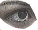
The white of the eye, the sclera, and the transparent part of the eye, the cornea, are basically the same kind of tissue. The cornea contains no blood vessels and is relatively dehydrated. The outside eyeball surface is bathed in tears, the inner surfaces of the cornea and the sclera are bathed by a clear fluid called aqueous humor. The iris is a disc behind the cornea with a variable opening. The opening is not under voluntary control. It is adjusted by the nervous system in response to the light intensity. The control is disabled by eye physicians when they try to see into the interior of the eye during an eye exam.
The back of the eye is coated with a complex,
light sensitive tissue called the retina. The
retina develops directly out of brain tissue and is
the first layer of the complex signal processing
system we possess. The processing starts at the
retina with its array of 120 million rods (sensors
for dim light) and about 6 million cones (sensors
for bright light.) CORRECTING IMPERFECT VISIONMore than half of the population have less than perfect vision. A "perfect" eye is able to form a
sharp image on the retina for object distances that
range from infinity (the stars) to a very short
distance from the eye (called the near point). Such
an eye is also capable of processing very dim light
(essentially one photon per receptor) to very
intense light (a bright, sun lit snow scene.) The
"perfect" eye can "see" two point sources as
distinct if the light rays from these sources for
an angle of one minute!! of arc at the eyeball. a
sharp image on the retina for object distances that
range from infinity (the stars) to a very short
distance from the eye (called the near point). Such
an eye is also capable of processing very dim light
(essentially one photon per receptor) to very
intense light (a bright, sun lit snow scene.) The
"perfect" eye can "see" two point sources as
distinct if the light rays from these sources for
an angle of one minute!! of arc at the eyeball.
 Two
"perfect" eyes must come with a perfect nervous
system which fuses the signals from the two retinas
into a single processed images which is perceived
to be three dimensional. Information from this
perceived image has to interact with the part of
the nervous system that controles movement so that
we can successfully reach for objects and walk
without bumping into things. Even such a perfect
system can be fooled by its own processing and
present us with various visual illusions. (They are
not optical illusions. The optics is OK, it's the
processing that is too simplistic and refuses to
accept the fact that your face is really not on the
other side of the mirror.) Two
"perfect" eyes must come with a perfect nervous
system which fuses the signals from the two retinas
into a single processed images which is perceived
to be three dimensional. Information from this
perceived image has to interact with the part of
the nervous system that controles movement so that
we can successfully reach for objects and walk
without bumping into things. Even such a perfect
system can be fooled by its own processing and
present us with various visual illusions. (They are
not optical illusions. The optics is OK, it's the
processing that is too simplistic and refuses to
accept the fact that your face is really not on the
other side of the mirror.) 
REFRACTIVE IMPERFECTIONS.The eye, as an optical system, has a range of focal distances which are determined by the curvature of the lens since the curvature of the cornea is fixed. In a normal eye the lens can change the effective focal length of the eye by as much as 15 diopters. This process is known as accomodation.MYOPIAIt is not uncommon that an eyeball comes equipped with a particular cornea-lens set which is too long. That is, the eyeball is longer than the longest attainable focal length. The idea of a "prefect" eyeball is that the retina is exactly at the focal plane of the cornea-lens optical system. That means that parallel ligth rays (coming from a far star) focus on the retina. If the eyeball
is longer than the longest attainable focal length,
then parallel light does not come to a focus at the
retina and the image of a far-away star is a disc
rather than a point. The star is seen as "blurred". If the eyeball
is longer than the longest attainable focal length,
then parallel light does not come to a focus at the
retina and the image of a far-away star is a disc
rather than a point. The star is seen as "blurred".
 As
the eye looks at closer objects the image distances
increase, the eye can handle that and the images
are sharp. Such eyes are called myopic or
nearsighted. Myopia can be corrected with diverging
lenses placed in front of the eye or by the
reshaping of the cornea. As
the eye looks at closer objects the image distances
increase, the eye can handle that and the images
are sharp. Such eyes are called myopic or
nearsighted. Myopia can be corrected with diverging
lenses placed in front of the eye or by the
reshaping of the cornea.Follow the links at the end of this narrative for more information. PRESBYOPIA To
see objects that are located close to the eye, the
focal length of the eye has to shorten. (Recall the
lens equation. To form a sharp image for various
values of the object distance and a fixed image
distance, f has to change.) Most people can squeeze
the lens enough to obtain sharp images for objects
that are about 10 inches away. Some myopes can even
do much better than that. As the eye ages, the
ability of the lens to flex diminishes. The eye
loses its power to accomodate. Since the lens loses
its ability to form short f's, the ability to see
close objects is lost. This is known as
presbyopia. To
see objects that are located close to the eye, the
focal length of the eye has to shorten. (Recall the
lens equation. To form a sharp image for various
values of the object distance and a fixed image
distance, f has to change.) Most people can squeeze
the lens enough to obtain sharp images for objects
that are about 10 inches away. Some myopes can even
do much better than that. As the eye ages, the
ability of the lens to flex diminishes. The eye
loses its power to accomodate. Since the lens loses
its ability to form short f's, the ability to see
close objects is lost. This is known as
presbyopia.The following chart shows how the abilty to adjust the focal length gets progressively worse with age. 
The horizontal axis represents the age, the vertical axis represents the range of diopters that the lens can add to the system. Note that at age 75 the eye is a fixed focal length system, good for only one object distance. If this used to be a "perfect" eye, the remaining object distance is infinity. If this is a myopic eye, the remaining object distance is the old far point. This chart by the way, is one of the most unbeatable tools to determine a person's age. There is no way to fake poor accomodation. Presbyopia is corrected with a set of converging lenses (or glasses.) Since the accomodation is at fault, different lenses are needed for different distances. A typical number is two. Reading glassed and distance glasses, or a two-part lens (a bifocal.) It is not uncommon to enecounter an eye that does not focus all parallel rays through the same point. Rays in different planes focus through different points. This condition is known as astigmatism. It is corrected by a lens that has a similar "defect" as the eye but in the other direction. Thanks to the Phys 251 students at IUPUI who donated eyes for this page.
You can get a lot more information about this subject on the internet. Here are a few search enginesAnd here are a few good links to get you started. The Anatomy of the Eye:1.  2. 3. 4. 2. 3. 4.
Correcting Vision: The Science of Vision Illusions Research Questions (1 point extra credit each!)
This site is made possible by
funding from the National Science Foundation
(DUE-9981111). |
 The
characteristic color of the eyes comes from the
pigment in the iris. The pigment from which the
iris gets its color is called melanin.
The
characteristic color of the eyes comes from the
pigment in the iris. The pigment from which the
iris gets its color is called melanin.  The is the
same pigment that colors our hair and skin. It is
the density and location of the melanin particles
that determine the "color" of the eye. Deep brown
eyes and light sky blue eyes use exactly the same
pigment.
The is the
same pigment that colors our hair and skin. It is
the density and location of the melanin particles
that determine the "color" of the eye. Deep brown
eyes and light sky blue eyes use exactly the same
pigment.
 These pigmented detectors enable us to distinguish
about 200 hues.
These pigmented detectors enable us to distinguish
about 200 hues.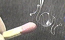 The last imperfection mentioned here has to do with
the transparency of the lens. For various reasons,
ranging from trauma to aging, the lens looses its
opacity as the tissue in the lens crystalizes. This
is known as cataract. The word cataract
means waterfall. Looking through such a lens
resembles looking through falling water. The
correction requires that the lens be removed.
People who had cataract surgery in the past had to
wear thick glassses. Today the original lens is
replaced by a polymer lens know as an intraocular
implant (IOL) shown on the left. The object to the
left of the lens is a wooden match.
The last imperfection mentioned here has to do with
the transparency of the lens. For various reasons,
ranging from trauma to aging, the lens looses its
opacity as the tissue in the lens crystalizes. This
is known as cataract. The word cataract
means waterfall. Looking through such a lens
resembles looking through falling water. The
correction requires that the lens be removed.
People who had cataract surgery in the past had to
wear thick glassses. Today the original lens is
replaced by a polymer lens know as an intraocular
implant (IOL) shown on the left. The object to the
left of the lens is a wooden match.